
Unveiling the Molecular Portals of Malaria How High-Resolution Imaging is Rewriting the Biology of Parasitic Invasion

The battle against malaria, a disease that has haunted human civilization for millennia, has entered a transformative new chapter as scientists finally visualize the microscopic "doors" used by parasites to hijack human blood cells. For decades, the exact mechanism by which the Plasmodium parasite breaches the defenses of a red blood cell remained one of biology’s most elusive mysteries. Now, leveraging the power of advanced cryo-electron microscopy, researchers have captured the first high-definition images of the "moving junction," a temporary molecular portal that the parasite constructs to slip into its host. This breakthrough, led by the lab of Chi-Min Ho at Columbia University, provides an unprecedented look at the biophysical maneuvers of one of the world’s most successful killers, offering a new roadmap for drug development.
The Ghost of Mala Aria: A Historical Context
To understand the weight of this discovery, one must look back at the long and lethal history of the disease. In medieval Italy, the illness was attributed to "bad air"—mala aria—rising from stagnant swamps. It was believed that miasmas, or foul vapors, were the direct cause of the fever, chills, and jaundice that decimated entire communities. It was not until 1880 that the French physician Charles Louis Alphonse Laveran, working in Algeria, identified single-celled protists in the blood of infected soldiers. He named the genus Plasmodium, a discovery that eventually earned him the Nobel Prize in 1907.
Despite identifying the culprit, the scientific community struggled for over a century to understand the mechanics of infection. Malaria remains a global health crisis; in the 20th century alone, it was responsible for up to 5% of all global deaths. Today, according to the World Health Organization, it continues to claim approximately 600,000 lives annually, with a disproportionate impact on children under the age of five in sub-Saharan Africa. The parasite’s ability to rapidly evolve resistance to frontline drugs like chloroquine and artemisinin has made the search for new biological vulnerabilities a matter of international urgency.
The Biophysics of a Microscopic Breach
The most critical stage of the malaria infection occurs when the parasite, in its "merozoite" form, encounters a human red blood cell. Red blood cells are among the smallest and most specialized cells in the body, measuring roughly 8 micrometers in diameter. The Plasmodium merozoite is a fraction of that size, yet it possesses a sophisticated toolkit for breaking and entering.
Previous research in the 1970s used early electron microscopy to observe a "thickening" of the cell membrane at the point of contact. However, the process was too fast and the structures too small for the technology of the era. The new research from the Ho lab has finally solved this puzzle. Using cryo-electron microscopy—a technique that freezes biological samples mid-action to preserve their natural state—the team imaged the "moving junction" in the very moment of invasion.
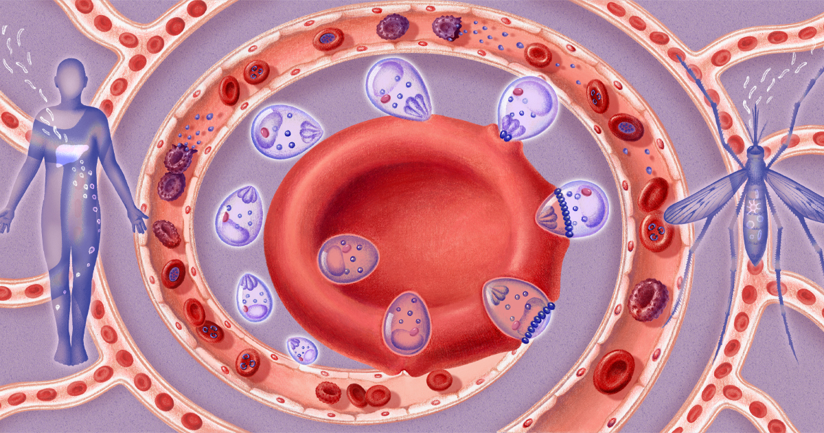
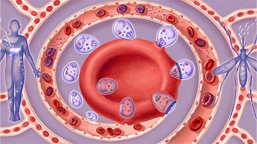
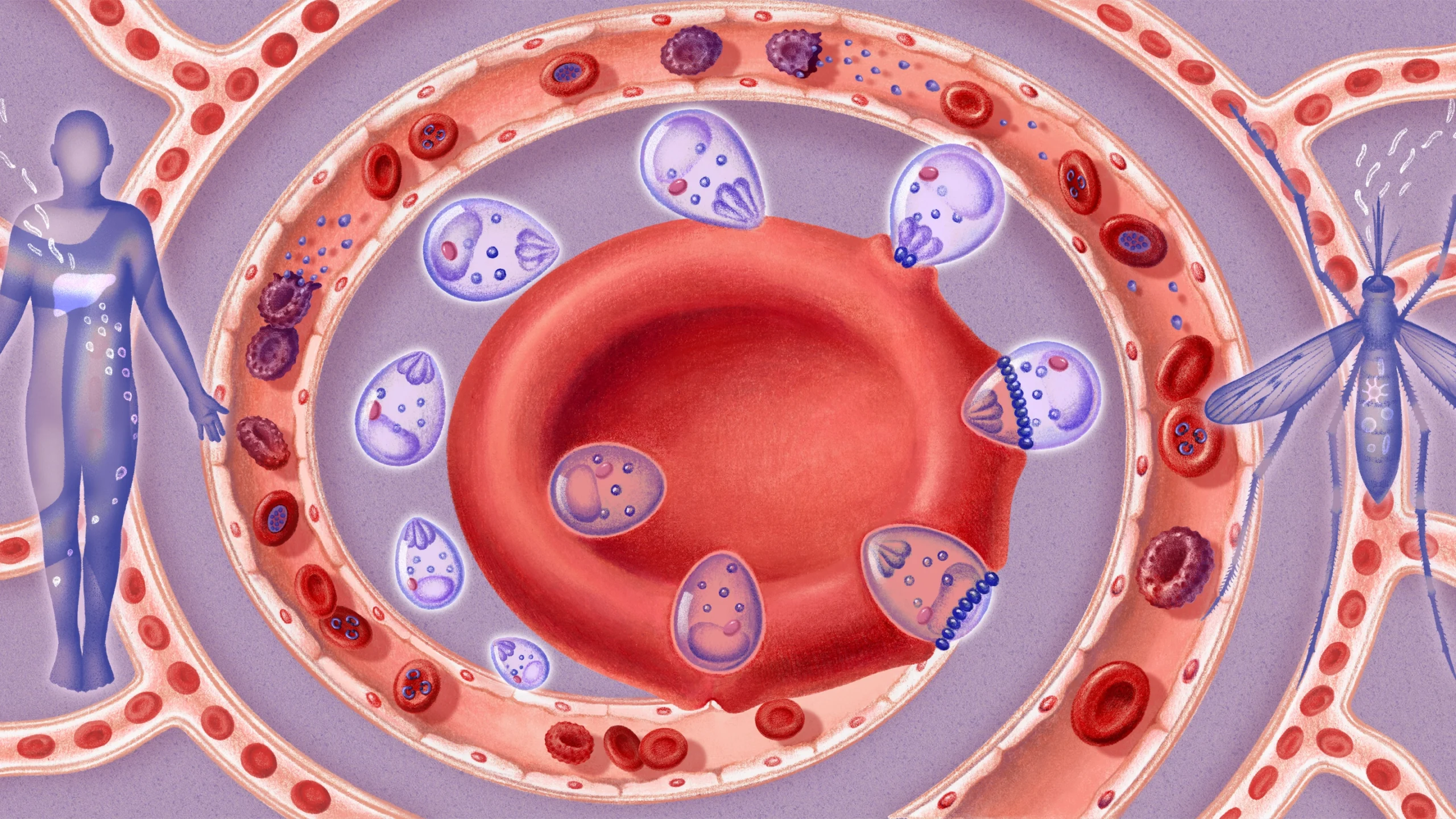
The moving junction is a ring-shaped structure that acts as a molecular gasket. As the parasite presses against the host cell, it injects a specific set of four protein units into the red blood cell’s membrane. Three of these proteins form a dense, wedge-like anchor beneath the surface, while a fourth protein acts as a tether, linking the parasite to the anchor. As the parasite pushes forward, these "anchors" create electrostatic forces that displace the lipid molecules of the host membrane.
Jake Baum, a parasitologist at the University of New South Wales, described the findings as a shift from traditional molecular biology to pure biophysics. The parasite does not simply "pop" the cell; it remodels the membrane, causing it to bow inward and eventually wrap around the invader. This ensures the host cell remains intact and functional, effectively turning the red blood cell into a protective bunker.
A Chronology of Infection: The 48-Hour Cycle
The invasion of the red blood cell is just the beginning of a complex and highly coordinated life cycle. Once the Plasmodium parasite has successfully navigated the moving junction, it begins a process of radical "home remodeling."
- Entry (Seconds): The moving junction forms and disappears in less than 60 seconds, leaving the cell membrane sealed and the parasite safely inside.
- Trophozoite Stage (Hours 1–24): The parasite transforms from a rigid, egg-shaped merozoite into a flexible, amoeba-like trophozoite. It begins to consume the host’s hemoglobin, the oxygen-carrying protein that gives blood its color.
- Remodeling (Hours 24–36): The parasite builds what researchers describe as "plumbing and flooring." It secretes proteins that stretch the cell’s cytoplasm and create transport vesicles to remove waste and import nutrients.
- Replication (Hours 36–48): The parasite undergoes asexual reproduction, creating dozens of new merozoites within the single host cell.
- Rupture: The host cell, now exhausted and packed with parasites, bursts open. The new merozoites flood the bloodstream, seeking out fresh red blood cells to repeat the cycle.
This rapid multiplication is why malaria symptoms can escalate so quickly. Within days of the initial infection, millions of parasites can be circulating in the host’s system, leading to the characteristic cycles of high fever and debilitating chills.
Evading the Spleen: The Sticker Strategy
One of the most fascinating aspects of the Plasmodium survival strategy is its ability to hide from the human immune system. The spleen serves as the body’s primary filter, identifying and removing damaged or stiffened red blood cells. An infected cell, filled with a growing parasite, would normally be a prime target for the spleen.
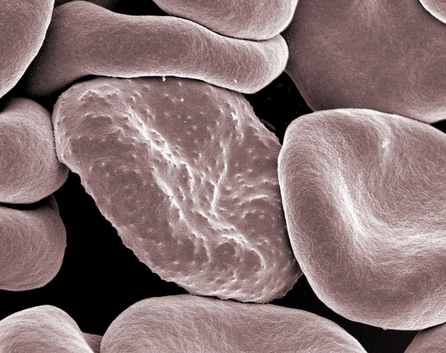
To avoid this fate, the parasite redecorates the exterior of the red blood cell with adhesive proteins. These proteins form small protrusions, or "knobs," on the cell surface. These knobs act like biological Velcro, causing the infected cells to stick to the walls of blood vessels. By anchoring themselves to the vascular endothelium, the parasites avoid being swept into the spleen.
However, this "sticker strategy" is also responsible for the disease’s most dangerous complications. When thousands of infected cells clump together in the small blood vessels of the brain or lungs, they cause blockages. This leads to cerebral malaria, organ distress, seizures, and comas. Research from Kirk Deitsch’s lab at Weill Cornell Medicine has further revealed that the parasite can periodically switch between different versions of these adhesive proteins, staying one step ahead of the immune system’s antibodies for weeks at a time.
Implications for Drug Discovery and AI Integration
The mapping of the moving junction provides a "choke point" that could be exploited by a new generation of antimalarial drugs. Historically, drug discovery for malaria was a process of trial and error—testing thousands of compounds to see which ones killed the parasite without knowing why.
"All that work was done essentially blind," noted Kirk Deitsch. With the structure of the moving junction now clear, researchers can move toward rational drug design. The Ho lab has already demonstrated the potential of this approach by using an AI-powered protein-design platform to create an "inhibitory protein." This synthetic molecule was designed to specifically block the moving junction proteins, preventing the parasite from ever entering the cell. In laboratory tests, the protein successfully stopped Plasmodium in its tracks.
However, significant hurdles remain. Because the invasion process lasts only about a minute, any drug targeting the moving junction must be incredibly fast-acting or have a very long half-life in the bloodstream. Furthermore, while the AI-designed protein worked in a test tube, its safety and effectiveness in the human body have yet to be determined.
The Broader Impact: A Skeleton Key for Parasitic Diseases
The significance of this research extends beyond malaria. Plasmodium belongs to a large phylum of parasites known as Apicomplexans. This group includes Toxoplasma gondii, which causes toxoplasmosis (a major concern for pregnant women and immunocompromised individuals), and Cyclospora, a common cause of foodborne illness.
Emerging evidence suggests that many of these parasites use nearly identical "invasion machinery" to enter host cells. By solving the structure of the Plasmodium portal, scientists may have found a "skeleton key" that could unlock treatments for a wide range of parasitic infections. Recent studies in 2026 have already begun to draw parallels between how Toxoplasma and Plasmodium sense the right moment to deploy their moving junctions, suggesting a conserved evolutionary mechanism across the phylum.
Conclusion: Chasing the White Whales
The high-resolution imaging of the malaria portal represents a milestone in structural biology, but for Chi-Min Ho and her colleagues, it is only the beginning. The lab is now turning its attention to other protein complexes involved in the infection cycle, including the adhesive proteins that allow infected cells to stick to blood vessel walls.
"We’ve caught one white whale," Ho said, referencing the long-sought structure of the moving junction. "So we want to go after the other remaining white whales."
As climate change continues to expand the range of malaria-carrying mosquitoes into new territories, the need for these "white whales" of research has never been more pressing. By combining biophysics, AI-driven design, and advanced microscopy, science is finally closing the doors that Plasmodium has used to devastate human health for centuries. The transition from "bad air" to molecular gaskets marks not just a victory for researchers, but a beacon of hope for the hundreds of millions of people living in the shadow of malaria.